Structure of the spinal column
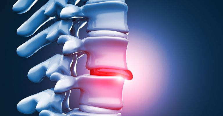
The spine has a segmental structure. To ensure its mobility, bony vertebral bodies alternate with cartilaginous intervertebral discs. The intervertebral discs are elastic enough to allow movement between the vertebrae. At the same time, however, they are also stable enough to withstand heavy loads. Intervertebral discs consist of the nucleus pulposus in the center and the annulus fibrosus, a fibrous ring around the outside of the nucleus pulposus. The intervertebral disc loses its elasticity under heavy strain or due to ageing. The gel-like core of the intervertebral disc bulges or tears. If the protrusion is so pronounced that it leads to compression in the area of the spinal cord, this is known as a herniated disc. This can lead to a temporary or even permanent loss of function of the nerve fibers on which the disc is pressing. This results in neurological deficits in theareas supplied by the affected nerve.
What is a slipped disc?
A herniated disc in dogs (disc prolapse) causes disc tissue to protrude from the gelatinous nucleus of the dog's intervertebral disc. The spinal column consists of bony vertebrae, some of which are fused together. The intervertebral discs lie between the bony vertebrae of the spine like a buffer. The intervertebral discs are made up of a ring-shaped, fibrocartilaginous tissue that surrounds a soft gelatinous core. In the event of a herniated disc in dogs, this gelatinous core protrudes from the disc and presses on the dog's spinal cord and/or surrounding nerves.
Symptoms
The symptoms caused by a herniated disc in dogs depend on the extent to which the disc protrudes into the spinal canal and where it occurs in the spine. Initially, there is usually only a protrusion of the intervertebral disc into the spinal canal. This is usually painful for the dog. Pain is often the first sign of disc disease. The dog's urge to move is often significantly restricted due to the pain. If the entire intervertebral disc tissue then prolapses (extrusion), the herniated disc in the dog manifests itself as paralysis of the front or hind legs with dragging of the affected limb, severe pain and increased
sensitivity to pain and pressure. In some cases, a stiff neck and an unnatural posture of the back, such as a hump, can also be signs of a herniated disc in dogs. If the disc tissue presses on the nerves responsible for controlling the bladder and anal sphincter muscles, this can result in incontinence in dogs.
Risk factors and causes
A herniated disc in dogs can be attributed to wear and tear of the disc tissue (degenerative change). This change can have several causes, for example incorrect loading or overloading of the spine, the dog being overweight or the normal ageing process. Herniated discs are therefore not uncommon in older dogs.
Some dog breeds suffer more frequently from slipped discs. For example, dogs with particularly short legs or a long back can develop a slipped disc even in middle age. On the one hand, the long back puts more strain on the intervertebral discs, and on the other hand, these breeds also tend to calcify early and thus lose their elasticity. As a result, the intervertebral discs lose their buffer function at an early stage.
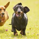
Slipped discs often occur in breeds with a long spine, such as dachshunds. This is why the disease is popularly known as "dachshund paralysis". However, other breeds can also suffer from herniated discs, such as the French Bulldog with its tendency to develop wedge vertebrae.
Type
A distinction is made between 2 types of herniated disc(Hansen's type 1 and type 2). They are the most common cause of degenerative damage to the spinal cord in dogs. Chondrodystrophic breeds in particular (e.g. Dachshunds, Pekingese) tend to develop early cartilaginous metaplasia of the nucleus pulposus with dystrophic calcification and consecutive protrusion (focal constriction in the spinal canal) or projectile-like prolapse of the nucleus pulposus into the spinal canal (extrusion; extensive spread of detritus in the epidural space). These changes, known as Hansen's type 1, are mainly localized in the thoracic and lumbar region. Hansen's type 2 occurs predominantly in non-chondrodystrophic, but also chondrodystrophic dog breeds. It rarely occurs in cats, horses and other species. Tears in the annulus fibrosus and fibrous metaplasia of the nucleus pulposus only occur in individual intervertebral discs, possibly induced by traumatic effects.
The direct ("primary injury") mechanical damage is followed by a heterogeneous spectrum of delayed tissue alterations ("secondary injury"). Patients often suffer from irreparable spinal neurological deficits with edema, axonopathy, dilated myelin sheaths and necrosis. Older spinal cord contusions may show hourglass-shaped atrophy.
Clinic
Disc herniations are more common in dogs than in cats. Dogs of chondrodystrophic breeds are overrepresented. In most cases there is degeneration of the disc, but explosive disc extrusions of healthy nucleus pulposus are also possible. Disc herniations most commonly occur in the thoracolumbar spine and cause mild to severe neurological deficits in the hindquarters depending on the induced spinal cord injury or compression. Cervical disc herniations can lead to severe tetraparesis or tetraplegia, but the symptoms are usually milder due to the wider rim around the spinal cord.
Diagnosis
The diagnosis is made using myelography, CT or MRI.
Therapy
Treatment can be conservative or surgical. Conservative treatment of an acute herniated disc is analogous to spinal cord trauma. Otherwise, analgesic therapy (NSAID, gabapentin) or treatment with steroids is primarily used. Strict immobilization must be observed. Surgical treatment usually involves a hemilaminectomy or mini-hemilaminectomy. The prognosis is often favourable, but depends on the severity of the clinical signs and the location of the lesion.
Treatment options
In the acute phase, physiotherapeutic treatment is usually not recommended due to the severe pain. Depending on recovery, careful stretching of the back and gentle massage can be started approximately 14 days after the acute phase. If the dog shows neurological deficits in one or more limbs, the affected limb can be treated directly while lying down. Suitable measures here are massage and reflex training.
Sources and relevant links
Schwarz S (2018). Physiotherapie für Hunde. 1. Auflage. Schattauer GmbH.
Patienteninformationen (2020). Version 1.0. Thieme.
Sigrist N (2022). Notfallmedizin für Hund und Katze. 2., aktualisierte Auflage. Thieme.
Baumgärtner W, Gruber A, Hrsg. Spezielle Pathologie für die Tiermedizin. 2., aktualisierte Auflage. Stuttgart: Thieme; 2020.
